Uncovering
The
Unseen
S.V. Paradoks Symposium 2025
Openingsspreker:
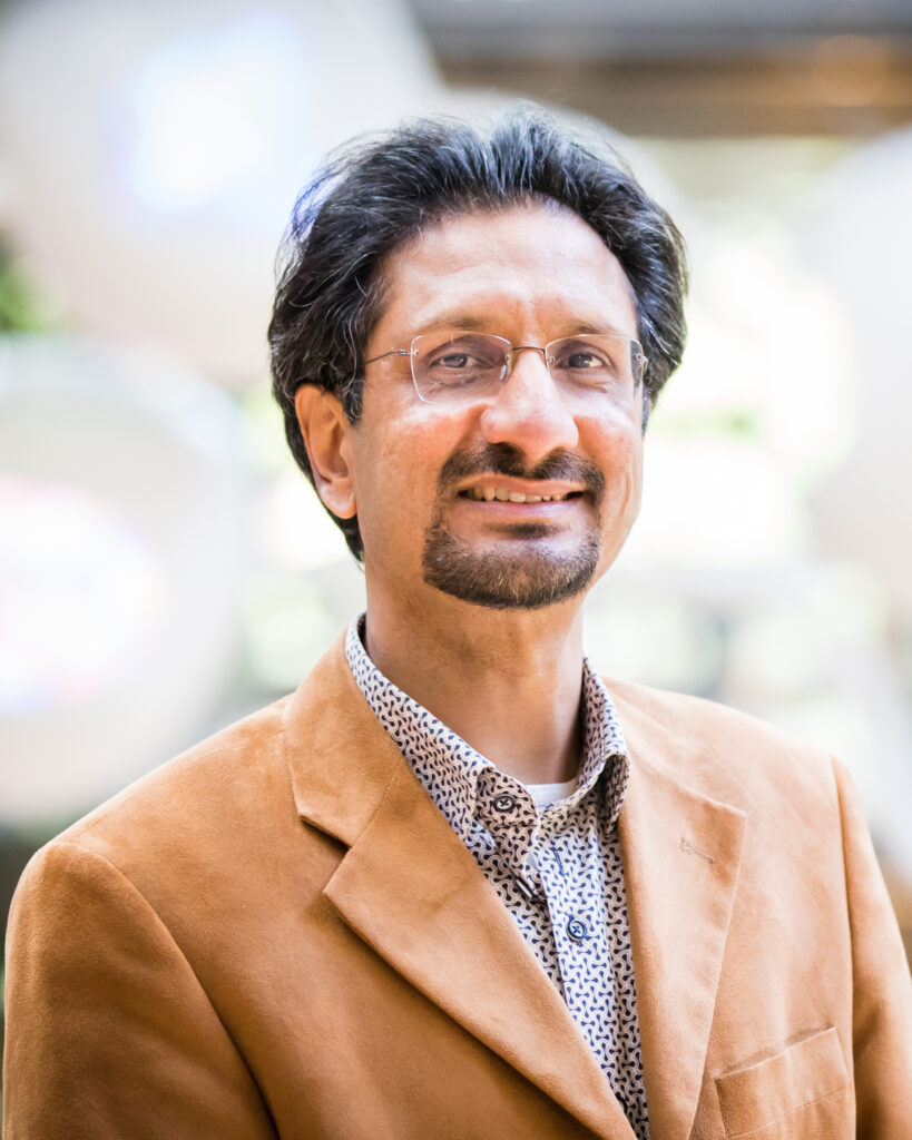
Srirang Manohar, Prof. Dr.
Multi-Modality Medical Imaging (M3I), TechMed Centre,
Universiteit Twente,
Nederland
Bloedsporen in het donker: borstkanker opsporen met geluid en licht
Wat als we verborgen sporen van borstkanker konden onthullen door bloed (en andere kenmerken) in kaart te brengen – zoals detectives bloedsporen opsporen en volgen? Met behulp van licht en geluid legt fotoakoestisch-ultrageluid tomografie de bloedvaten diep in de borst bloot als nooit tevoren en kan het helpen borstkanker in een vroeg stadium te detecteren. Tijdens deze lezing nemen we u mee in de basisprincipes van de fysica, de in Twente ontwikkelde beeldvormingstechnologie en de mogelijke klinische toepassingen voor borstkankerdiagnostiek. Daarnaast zal er een demonstratie zijn waarin we het effect achter de fotoakoestische methode laten zien.
Sprekersbiografie
Srirang Manohar is hoogleraar en voorzitter van de onderzoeksgroep Multi-Modality Medical Imaging (M3I) aan de Universiteit Twente. Hij is gespecialiseerd in niet-invasieve diagnostische technologieën zoals fotoakoestische en echografische beeldvorming. Hij heeft leiding gegeven aan PAMMOTH, een Europees project gericht op de ontwikkeling van een stralingsvrij en real-time diagnostisch apparaat voor borstkanker. Zijn onderzoek richt zich ook op schildklierproblemen, prostaatkanker en reumatoïde artritis. Srirang combineert de ontwikkeling van innovatieve technologieën met vroege klinische toepassingen om diagnostische en therapeutische resultaten te verbeteren.
English
Srirang Manohar, Prof. Dr.
Multi-Modality Medical Imaging (M3I), Tech Med Centre,
University of Twente,
The Netherlands
Bloodtracks in the dark: uncovering breast cancer with sound and light
What if we could reveal hidden traces of breast cancer by tracking blood (amongst other features) – the way detectives uncover and follow bloodstains? Using light and sound, photoacoustic-ultrasound tomography exposes the blood vessels deep in the breast as never before and may help us uncover breast cancer at its earliest stages. Join me as we go through the basic physics, the home-grown Twente imaging technology, and its potential clinical application of breast cancer imaging. We will also have a demonstration that shows the effect behind the method of photoacoustics.
Speaker biography
Srirang Manohar is Professor and Chair of Multi-Modality Medical Imaging (M3I) at the University of Twente, specializing in non-invasive diagnostic technologies like photoacoustic and ultrasound imaging. He has lead PAMMOTH, a European project to develop a radiation-free and real-time breast cancer diagnostic device. His research also targets thyroid issues, prostate cancer and rheumatoid arthritis. Srirang combines novel technology development with early clinical applications to improve diagnostic and therapeutic outcomes.
Ronde 2:
Technologie aan de voorhoede
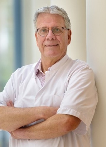
Theo Ruers studeerde in 1985 af aan de Faculteit Geneeskunde in Maastricht. In 1989 behaalde hij zijn PhD in de immunologie. Hij werd opgeleid tot chirurg in Maastricht en Eindhoven en specialiseerde zich vervolgens twee jaar in de oncologische chirurgie aan het Kings College Hospital en het Hammersmith Hospital in Londen (1992-1994). Na functies als oncologisch chirurg in Maastricht en Nijmegen werd hij in 2007 hoofd van de afdeling Chirurgische Oncologie bij het Nederlands Kanker Instituut in Amsterdam. In 2010 werd hij benoemd tot klinisch hoogleraar aan de Technische Universiteit Twente, waar hij zich richt op de toepassing van biomedische beeldvorming en behandelstrategieën in de oncologie.
In de klinische praktijk richt hij zich voornamelijk op darmkanker en leverchirurgie, met een bijzondere interesse in beeldgestuurde chirurgische ingrepen. Theo Ruers leidt een onderzoeksafdeling die zich bezighoudt met beeldvormingstechnieken voor de diagnostiek en behandeling van kanker. Dit onderzoek richt zich op onderwerpen zoals elektromagnetische chirurgische navigatie, optische weefselanalyse met behulp van diffuse reflectiespectroscopie en hyperspectrale beeldvorming. Zijn doel is om chirurgische ingrepen te optimaliseren door de introductie van technologie en AI in de operatiekamer, wat leidt tot betere chirurgische en patiëntuitkomsten.
Hij is lid van verschillende internationale verenigingen voor chirurgische oncologie en onderzoeksnetwerken. Momenteel leidt hij het instituut voor vroege kankeropsporing bij het Nederlands Kanker Instituut.
English
Technology at the cutting edge
Theo Ruers graduated in 1985 from the Faculty of Medicine in Maastricht. He received his PhD in immunology 1989. He was trained as surgeon in Maastricht and Eindhoven and later for 2 years he specialised in surgical oncology at Kings College Hospital and the Hammersmith Hospital in London (1992-1994). After a position as surgical oncologist in Maastricht and Nijmegen, he became head of the Division Surgical Oncology at The Netherlands Cancer Institute Amsterdam (2007). He was appointed in 2010 as clinical professor at the Technical University of Twente on the application of biomedical imaging and treatment strategies in oncology.
In clinical practice his main field of interest is colorectal cancer and liver surgery, with special interest for image guided surgical procedures. Theo Ruers is heading a research division focussed on imaging techniques for diagnosis and treatment of cancer. This research concentrates around topic of electromagnetic surgical navigation, optical tissue diagnosis by diffuse reflectance spectroscopy and hyperspectral imaging. His focus is to optimize surgical interventions by the introduction of technology and AI in the OR, leading to better surgical and patient outcome. He is a member of several international surgical oncology societies and research societies. Currently he is leading the institute for early cancer detection at the Netherlands Cancer Institute
Casus
Finapres heeft de toonaangevende technologie voor continue niet-invasieve bloeddrukmeting en hemodynamica! Finapres ontwikkelt en distribueert medische systemen en software voor continue niet-invasieve hemodynamische monitoring. Deze systemen zijn het resultaat van meer dan 30 jaar onderzoek en gebruikerservaringen op dit vakgebied. De markten waarin Finapres actief is, zijn onderzoek (van ruimtevaart tot slaapstudie), klinische diagnostiek (autonome dysfunctie, cardiovasculair ziekten) en onderwijs.
Een potentiële nieuwe doelgroep voor Finapres zijn patiënten met tremor (bijvoorbeeld Parkinson) omdat deze groep naast motorische symptomen ook vaak klachten van autonome dysfunctie hebben. Vragen waar wij nog geen antwoord op hebben, zijn: Is het mogelijk om bij deze patiënten bloeddruk te meten met de huidige technologie van Finapres? Zo niet, zijn er oplossingen te bedenken waarbij deze patiënten wel gemeten kunnen worden met de huidige technologie?
English
Case
Finapres is the leading provider of technology for continuous non-invasive blood pressure measurement and hemodynamics! Finapres develops and distributes medical systems and software for continuous non-invasive hemodynamic monitoring. These systems are the result of over 30 years of research and user experience in this field. The markets in which Finapres operates include research (ranging from aerospace to sleep studies), clinical diagnostics (autonomic dysfunction, cardiovascular diseases), and education.
A potential new target group for Finapres includes patients with tremor (e.g., Parkinson’s disease), as this group often experiences autonomic dysfunction in addition to motor symptoms. However, there are still questions we need to answer: Is it possible to measure blood pressure in these patients using Finapres’ current technology? If not, what solutions could be developed to enable accurate measurement for these patients using the existing technology?
Ronde 3:
SpatialOmics: Technologie van cel naar chirurg
Prof. Dr. Ron M.A. Heeren behaalde in 1992 een PhD in de technische natuurkunde aan de Universiteit van Amsterdam. Hij begon zijn werk op het gebied van moleculaire beeldvorming en instrumentontwikkeling als onderzoeksgroepleider bij FOM-AMOLF in Amsterdam. Van 2001 tot 2019 was hij hoogleraar aan de faculteit Scheikunde van de Universiteit Utrecht, terwijl hij de beeldvormingsgroep bij AMOLF leidde.
In 2014 werd hij benoemd tot Distinguished Professor en Limburg Chair aan de Universiteit Maastricht. Hij is de oprichter en wetenschappelijk directeur van M4I, het Maastricht MultiModal Molecular Imaging Institute op de Brightlands Maastricht Health Campus. In 2021 werd hij verkozen tot lid van de Koninklijke Nederlandse Akademie van Wetenschappen (KNAW). Sinds 2024 is hij ambassadeur van de Technische Universiteit van München.
Zijn academische onderzoek richt zich op massaspectrometrie-gebaseerde gepersonaliseerde geneeskunde, translationele moleculaire beeldvorming en “omics”-onderzoek, high-throughput bioinformatica en de ontwikkeling en validatie van innovatieve moleculaire analytische beeldvormingstechnieken binnen verschillende wetenschappelijke disciplines.
English
Translational molecular imaging: technology from cell to surgeon
Prof. Dr. Ron M.A. Heeren obtained a PhD degree in technical physics in 1992 at the University of Amsterdam. He started to work on molecular imaging instrumentation and its application as a research group leader at FOM-AMOLF, Amsterdam. From 2001-2019, he was professor at the chemistry faculty of Utrecht University while running the imaging group at AMOLF. In 2014 he started as distinguished professor and Limburg Chair at Maastricht University. He is the founder and scientific director of M4I, the Maastricht MultiModal Molecular Imaging institute on the Brightlands Maastricht Health campus. In 2021 he was elected as a member of the Royal Dutch Academy of Sciences, KNAW. He has been an ambassador of the Technical University of Munich since 2024. His academic research interests are mass spectrometry based personalized medicine, translational molecular imaging and “omics” research, high-throughput bioinformatics and the development and validation of innovative molecular analytical imaging techniques across the scientific disciplines.
De toekomst van MRI
Hoewel conventionele MRI als duur en complex wordt beschouwd, zal prof. Klomp een tegenovergestelde visie presenteren. Door gebruik te maken van innovatieve concepten in supergeleiding, ultrasone systeemwerking en volledige automatisering van patiëntbeheer met AI-diagnostiek, wordt een nieuwe vorm van zelfscanning geïntroduceerd.
Prof. Klomp heeft een onderzoekslijn opgezet met meer dan 75 onderzoekers, gericht op uiterst precieze structurele en metabole MRI met ultra-hoge veldsterktes. Sinds vorig jaar leidt hij het onderzoek naar Beeldvorming en Oncologie (>350 onderzoekers) aan het UMC Utrecht, waar hij onder andere het eerste Scan2Go-concept ter wereld heeft gelanceerd in een extramurale ouderenzorginstelling (QuaRijn).
English
The future of MRI
While conventional MRI is considered expensive and complex, prof Klomp will reveal the opposed perception. Utilizing novel concepts in super conductivity, apply ultrasonic system operation and fully automate patient management with AI diagnosing, a next level of self-scanning will be shown. Prof Klomp setup a research line of over 75 researchers facilitating high precision structural and metabolic MRI using ultra high fields and since last year is managing research of Imaging and Oncology (>350 researchers) at the UMC Utrecht where a.o. he launched world’s first Scan2Go concept in an out centered elderly care facility (QuaRijn).
Ronde 4:

Revolutie in Gecombineerde Moleculaire & Structurele Beeldvorming: Ontwikkeling en Toepassing van een Veelzijdig Multimodaal Platform voor Theranostische en Multiplex Optische, Nucleaire en Anatomische Beeldvorming
Prof. Freek Beekman (1961, Twente) is wetenschapper en ondernemer in de life sciences. Hij leidt de sectie Biomedische Beeldvorming aan de TU Delft en is oprichter van MILabs (www.milabs.com) en oprichter/CEO van Free Bee Int’l (www.freebeeint.com). Zijn onderzoeksinteresses omvatten de ontwikkeling van detectoren en beeldreconstructie-algoritmen voor Positron Emissie Tomografie (PET), Single Photon Emission Computed Tomography (SPECT), Optische Tomografie en Röntgen CT, hybride fotonische beeldvormingsapparaten en de toepassing van AI op beeldvorming.
De kleine dier-fotonische systemen die door Freek en zijn teams bij TU Delft en MILabs zijn ontwikkeld, worden wereldwijd gebruikt in toonaangevende biomedische onderzoeksinstellingen, waaronder academische centra en grote farmaceutische bedrijven. Deze systemen hebben geleid tot talrijke wetenschappelijke ontdekkingen en dragen bij aan de ontwikkeling van nieuwe tracers en geneesmiddelen.
Deze presentatie richt zich op de ontwikkeling en toepassing van een geïntegreerd beeldvormingsplatform dat de unieke mogelijkheden van ultra-hoge resolutie PET, SPECT, Optische Tomografie en Röntgen CT combineert en optimaliseert.
English
Revolutionizing Combined Molecular & Structural Imaging: Development and application of a Versatile Multimodal Platform for Theranostic and Multiplexed Optical, Nuclear, and Anatomical Imaging
Prof. Freek Beekman (1961, Twente) is a scientist and life science entrepreneur. He heads the section Biomedical Imaging at TU Delft and is founder of MILabs (www.milabs.com) and founder/CEO of Free Bee Int’l (www.freebeeint.com). His research interests include development of detectors, image reconstruction algorithms for Positron Emission Tomography (PET), Single Photon Emission Computed Tomography (SPECT) Optical Tomography and X-ray CT, hybrid photonic imaging devices and AI applied to imaging. Today the small animal photonic systems that are developed by Freek and his teams at TU Delft and MILabs are in use in many premier biomedical research institutions worldwide including academic institutions and the largest blue chip pharma companies. These systems have resulted in many interesting discoveries and support the development of new tracers and pharmaceuticals. This presentation is about the development and application of an integrated imaging platform that synergizes (unique) capabilities of ultra-high-resolution PET, SPECT, Optical Tomography and X-ray CT.
Uncovering ultrasound to image the unseen: ultra-fast and super-simple
Chris L. de Korte is hoogleraar Medische Ultrasound Beeldvorming (zowel aan het Radboudumc als aan de Universiteit Twente) en ontwikkelt al meer dan 30 jaar innovatieve echografietechnieken. Hij is een autoriteit op het gebied van beeldvorming van functionele eigenschappen van weefsels met elastografie en flow-imagingtechnieken. Zijn klinische focus ligt op cardiovasculaire ziekten en borstkanker.
In de afgelopen tien jaar heeft hij AI-gestuurde handheld echografieapparaten ontwikkeld voor prenatale diagnostiek in omgevingen met beperkte middelen, evenals voor de detectie van heupdysplasie, artrose, aorta-aneurysma’s en andere aandoeningen in de huisartsenpraktijk en op consultatiebureaus in Nederland. Tijdens zijn presentatie zal hij de brug slaan tussen geavanceerde ultrasnelle 3D-echografie en eenvoudig te gebruiken handheld apparaten voor zorgprofessionals met minimale training.
Chris studeerde Elektrotechniek aan de Technische Universiteit Eindhoven en behaalde in 1999 zijn PhD aan de Erasmus Universiteit Rotterdam. In 2002 trad hij toe tot het Klinisch Fysisch Laboratorium van de afdeling Kindergeneeskunde van het Radboudumc, waarvan hij in 2004 hoofd werd. Sinds 2012 is hij voorzitter van het Medical UltraSound Imaging Centre binnen de afdeling Medische Beeldvorming: Radiologie van het Radboudumc. In 2015 werd hij benoemd tot hoogleraar Medische Ultrasound Beeldvorming aan het Radboudumc en in 2016 aan de Universiteit Twente.
Voor zijn onderzoek ontving hij meerdere subsidies van de Nederlandse Technologie Stichting (STW) en een VENI (2000), VIDI (2006) en VICI (2011) beurs van de Nederlandse Organisatie voor Wetenschappelijk Onderzoek (NWO). In 2018 ontving een consortium onder zijn leiding de NWO-TTW Perspectief-subsidie (4 miljoen euro). Hij is voorzitter van de Nederlandse Vereniging voor Medische Ultrasound (NVMU) en General Chair van het IEEE International Ultrasonics Symposium 2025, dat in september 2025 in Utrecht zal plaatsvinden.
English
Uncovering ultrasound to image the unseen: ultra-fast and super-simple.
Chris L. de Korte is Full Professor Medical Ultrasound Imaging (both at Radboudumc and at the University of Twente) and has developed innovative ultrasound techniques for more than 30 years. He is an authority in the field of imaging functional properties of tissues by elastographic and flow imaging techniques. His clinical focus is on cardiovascular diseases and breast cancer. In the last 10 years, he developed AI driven handheld ultrasound devices for prenatal diagnosis in low resource settings as well as in the field of hip dysplasia, osteoarthritis, aortic aneurism detection etc. for general practitioners and childcare centres in the Netherlands. In the presentation he will bridge the gap from high-end ultrafast 3D ultrasound to super simple handheld devices that can be used by health professionals with minimal training.
Chris studied Electrical Engineering at the Eindhoven University of Technology. In 1999, he obtained his Ph.D ) Erasmus University Rotterdam). In 2002 he joined the Clinical Physics Laboratory, Department of Paediatrics of the Radboud University Nijmegen Medical Center of which he became head in 2004. Since 2012, he is chair of the Medical UltraSound Imaging Centre at the Department of Medical Imaging: Radiology of Radboud university medical centre. He was appointed full professor Medical Ultrasound Imaging at Radboudumc (2015) and the University of Twente (2016). For his research he received multiple grants from the Dutch Technology Foundation (STW) and a VENI (2000), VIDI (2006) and VICI (2011) grant from The Netherlands Organisation for Scientific Research (NWO). In 2018, a consortium under his leadership was awarded the NWO-TTW Perspectief grant (4 Meuros). He is president of the Netherlands Society for Medical Ultrasound (NVMU) He is the General Chair of the 2025 IEEE International Ultrasonics Symposium that will take place in September 2025 in Utrecht, The Netherlands.

Ronde 5:
Fluorescentie Imaging in het MST
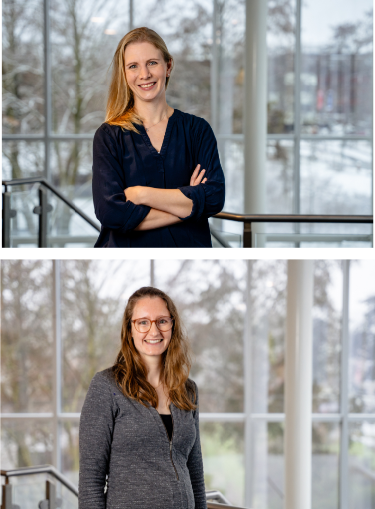
Fluorescentie imaging (FI) is een breed inzetbare techniek die operaties effectiever en veiliger kan maken, maar voor goed gebruik is ervaring en inzicht nodig. In Medisch Spectrum Twente is het FI lab opgericht om de technologische kennis te bundelen en eindgebruikers uit de gehele kliniek te ondersteunen. Hier zijn twee Technisch Geneeskundigen actief: Harry Vaassen en Anke Christenhusz.
Harry is betrokken sinds de oprichting van het FI lab. Sinds zijn afstuderen heeft hij onderzoek gedaan naar het gebruik van FI voor de beoordeling van darmdoorbloeding op de operatiekamer. Hij zal vertellen over de brede inzet van FI in MST en het nut van een FI lab.
Anke heeft zich tijdens haar promotietraject gespecialiseerd in de inzet van nieuwe technologie bij de behandeling van borstkanker. Na haar promotie heeft ze zich gevoegd bij het FI lab in MST, om de implementatie van FI en andere innovaties verder vorm te geven.
English
Fluorescence Imaging at MST
Fluorescence imaging (FI) is a highly versatile technique that can make surgeries more effective and safer. However, proper use requires experience and insight. At Medisch Spectrum Twente (MST), the FI lab was established to centralize technological knowledge and support end users across the clinic. Two Technical Physicians, Harry Vaassen and Anke Christenhusz, are actively involved in this initiative.
Harry has been involved since the founding of the FI lab. Since graduating, he has conducted research on the use of FI for assessing intestinal perfusion in the operating room. He will discuss the broad applications of FI at MST and the value of an FI lab.
Anke specialized in the use of new technology for breast cancer treatment during her PhD research. After completing her PhD, she joined the FI lab at MST to further develop the implementation of FI and other innovations.
Toveren met 3D medische beelden door Rob van Doremalen
In het medische 3D lab van MST gebruiken we medische beelden om anatomie of pathologie van de patiënt inzichtelijker te maken, gewenste situatie te simuleren of zelf patiënt specifieke hulpmiddelen te ontwikkelen. Dit doen we voor verschillende medische vakgroepen en veelal gebruiken we hiervoor medische beeldvorming die voor diagnostiek al beschikbaar is. Het team bestaat uit meerder technisch geneeskundigen, medisch technici, een klinisch fysicus, quality officer en een teamhoofd. Rob van Doremalen is één van de technisch geneeskundigen sinds 2021. Hij is in 2015 afgestudeerd in Twente, heeft daarna een jaar in het 3D lab van Radboudumc gewerkt. Vervolgens heeft hij een promotietraject doorlopen in Twente samen met de ziekenhuizen ZGT en Deventer Ziekenhuis, afgerond in 2021, met focus op hoe 3D technieken in de zorg het best ingezet kunnen worden. Parallel is Rob innovatie coördinator geweest in Deventer Ziekenhuis en sinds een jaar programma manager van het InnovatieLab in MST. Tijdens deze sessie zal Rob een aantal voorbeelden laten zien hoe ze impact hebben op de behandeling van patiënten door medische beelden anders weer te geven en hoe ze georganiseerd zijn in MST.
English
Magic with 3D Medical Imaging
In the medical 3D lab at MST, we use medical imaging to provide better insight into patient anatomy or pathology, simulate desired situations, and even develop patient-specific medical devices. We do this for various medical specialties, often utilizing imaging that is already available for diagnostics.
The team consists of multiple technical physicians, medical engineers, a clinical physicist, a quality officer, and a team leader. Rob van Doremalen has been one of the technical physicians since 2021. He graduated from Twente in 2015 and spent a year working at the 3D lab of Radboudumc. He then pursued a PhD in Twente in collaboration with ZGT and Deventer Hospital, completing it in 2021, focusing on the optimal integration of 3D technologies in healthcare. Alongside his PhD, Rob was an innovation coordinator at Deventer Hospital and has been the program manager of the Innovation Lab at MST for the past year.
During this session, Rob will showcase several examples of how they enhance patient treatment by presenting medical images in new ways and how their work is organized within MST.
Ronde 6:
IJzer Nanodeeltjes-MRI: Cel-Specifieke Detectie van kleine Metastasen, MS en Plaques
Jelle Barentsz behaalde in 1980 zijn artsexamen. Hierna vervulde hij zijn militaire dienstplicht als arts-assistent op de röntgenafdeling van het Militair Hospitaal te Utrecht. In 1985, na zijn specialisatie in Nijmegen, werd hij stafmedewerker aan de afdeling Radiologie van het Radboudumc. In 1990 promoveerde hij en in 1998 werd hij tot hoogleraar Functionele Beeldvorming benoemd. Hij richtte in 2011 het Radboudumc Prostaat Expertise Centrum op.
De wetenschappelijke focus van Barentsz ligt op het gebied van prostaatkanker- en lymfeklierbeeld-vorming. Met zijn team ontwikkelt en implementeert hij nieuwe MRI-technieken. Hij zet zich met name in om de resultaten van zijn wetenschappelijk onderzoek in de dagelijkse praktijk toe te passen. Zijn belangrijkste bijdragen zijn: MRI-onderzoek met nano-vloeistof voor het opsporen van kleine lymfekliermetastasen, ontwikkeling en implementatie van prostaat-MRI en het PI-RADS-scoresysteem voor de gestandaardiseerde interpretatie van prostaat-MRI’s.
English
Iron Nanoparticle MRI: Cell-Specific Detection of Small Metastases, MS, and Plaques
Jelle Barentsz obtained his medical degree in 1980. He then fulfilled his military service as an assistant physician in the radiology department of the Military Hospital in Utrecht. After completing his specialization in Nijmegen in 1985, he became a staff member at the Radiology Department of Radboudumc. In 1990, he obtained his PhD, and in 1998, he was appointed Professor of Functional Imaging. In 2011, he founded the Radboudumc Prostate Expertise Center.
Barentsz’s scientific focus is on prostate cancer and lymph node imaging. Together with his team, he develops and implements new MRI techniques and is particularly committed to translating his research into daily clinical practice. His key contributions include:
- MRI with nano-fluid for detecting small lymph node metastases
- Development and implementation of prostate MRI
- Creation of the PI-RADS scoring system for standardized interpretation of prostate MRIs

contact
S.V. Paradoks
Universiteit Twente
Hallenweg 5
Technohal
7522 NH Enschede
Met trots aangedreven door WordPress